The temporomandibular joint is one of the most complex articulations in the human body. On each side of the skull, a mandibular condyle articulates with the temporal bone via an articular disc — a biconcave fibrocartilaginous cushion — held in dynamic relationship by the lateral pterygoid muscle, the joint capsule, and a network of ligaments. During jaw opening, the condyle translates forward along the articular eminence; during chewing, it rotates and glides simultaneously in three dimensions. When any component of this system is overloaded, displaced, or inflamed, the result is temporomandibular dysfunction.
Epidemiological data suggest that 20–30% of adults will experience clinically significant TMD symptoms at some point in their lives, with peak prevalence in the 20–40 age group and a higher incidence in women. Despite this prevalence, TMD frequently goes undiagnosed — or is misattributed to sinus problems, ear pathology, or primary headache disorders — because its symptom profile overlaps significantly with other conditions.
Prosthodontists are uniquely placed to assess and manage TMD. Our specialty centres on occlusion — the way upper and lower teeth relate during contact and jaw movement — and on the interface between dental structures and jaw-joint function. Where a general dentist treats teeth in isolation, a prosthodontist evaluates the entire masticatory system: joint, muscles, nerves, teeth, and how prosthetic restorations affect or can correct bite discrepancies that drive TMD.
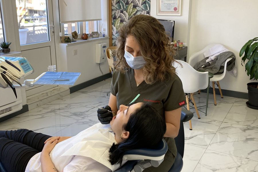
TMJ consultation — Dr. Dt. Tuba Akcakus Battal, prosthodontic specialist
Causes of TMJ Disorders
Bruxism and Parafunctional Habits
Bruxism — the involuntary grinding or clenching of teeth — is among the most common causes of TMD. Sleep bruxism, classified as a sleep-related movement disorder, generates bite forces far exceeding those of normal chewing: studies using electromyography have recorded masticatory muscle activity during sleep bruxism at 60–80% of maximum voluntary contraction. These forces are transmitted directly to the TMJ, compressing the articular disc and fatiguing the lateral pterygoid. Awake bruxism, by contrast, is typically clenching rather than grinding and is strongly associated with psychological stress.
Parafunctional habits extend beyond grinding: habitual nail-biting, pen-chewing, prolonged gum chewing, and resting the chin on the hand all impose asymmetric loading on the TMJ. Over time, masseter hypertrophy — a visible enlargement of the jaw muscle at the angle of the mandible — develops in chronic bruxers, and its own increased resting tone perpetuates joint compression even without active grinding.
Malocclusion and Bite Discrepancies
An uneven bite is not always the primary cause of TMD, but it is frequently a significant contributing factor — particularly when malocclusion forces the mandible into an adapted, displaced position to achieve maximum intercuspation. Deep overbite, posterior crossbite, and unilateral missing teeth (causing the jaw to shift laterally on closure) can all alter condylar position within the fossa. Poorly fitting prosthetics — over-contoured crowns, incorrect vertical dimension in dentures or implant-supported restorations — similarly disturb the occlusal equilibrium and can precipitate or worsen TMD symptoms.
Trauma and Stress
Direct trauma to the mandible or condyle — from a fall, road traffic accident, or sports injury — can cause condylar fracture, haemarthrosis, or initiate disc displacement that manifests as TMD weeks or months after the original injury. Indirect trauma, such as the hyperextension–flexion mechanism of whiplash, has also been associated with the onset of TMD through cervical muscle guarding that loads the TMJ asymmetrically.
Psychological stress acts through a different pathway: elevated cortisol and sympathetic nervous system activation increase masticatory muscle tone and parafunctional activity, while simultaneously lowering the pain threshold in the trigeminal system. Forward head posture — prevalent in sedentary, screen-based work — mechanically increases compressive load on both the cervical spine and the TMJ, and is an underappreciated postural driver of chronic jaw pain.
Articular Disc Displacement
The articular disc can be displaced from its normal position anterior to the condyle, most commonly due to laxity of the posterior attachment (bilaminar zone) caused by trauma or sustained joint compression. In disc displacement with reduction, the disc snaps back onto the condyle during opening, producing the characteristic clicking or popping sound at a reproducible point in the range of motion. In disc displacement without reduction, the disc remains anteriorly displaced and acts as a mechanical block, resulting in limited and deflected mouth opening — the classic "closed lock" presentation. Advanced untreated disc displacement may progress to disc perforation and direct bone-on-bone articulation, producing crepitus (a grinding, grating sound) and degenerative joint changes detectable on CBCT or MRI.
Symptoms of TMD
- Jaw pain or tenderness, particularly on waking or after meals
- Clicking, popping, or grating sounds on jaw movement
- Headaches — often frontal or temporal, mimicking tension headache or migraine
- Ear pain and tinnitus (referred pain from the TMJ via shared innervation)
- Limited mouth opening or jaw locking (acute closed lock: opening <30 mm)
- Facial muscle fatigue and soreness, especially in the masseter and temporalis
- Neck and shoulder tension (myofascial spread of masticatory muscle hyperactivity)
- Tooth sensitivity without caries (from grinding forces causing dentinal stress)
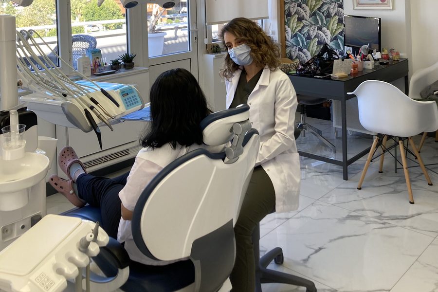
Dr. Dt. Tuba Akcakus Battal — TMJ clinical consultation, Antalya
Diagnosis
Clinical Examination
A systematic TMD examination begins with palpation of the masticatory muscles — masseter (body and origin), temporalis (anterior, middle, and posterior bellies), and lateral pterygoid (accessed intra-orally via the pterygomandibular space) — recording tenderness on a 0–3 scale. Maximum mouth opening is measured with a millimetre ruler between the upper and lower central incisors; a normal finding is ≥40 mm. Reduced opening (<35 mm), deviation of the mandible on opening, and deflection (persistent lateral shift that does not correct) are all diagnostically significant. The condylar loading test — applying upward and forward force to the mandible while the patient bites on a wooden spatula on one side — helps localise true intra-articular pain versus muscle pain. Finally, joint sounds are auscultated and palpated bilaterally during opening, closing, and lateral excursions.
Imaging
The dental panoramic radiograph (OPG) is first-line imaging for TMD: it provides an overview of condylar morphology, identifies gross degenerative changes (flattening, erosion, osteophyte formation), and rules out other pathology such as condylar hyperplasia or neoplasm. When condylar bone changes are suspected in more detail, cone beam computed tomography (CBCT) provides three-dimensional hard-tissue assessment with lower radiation dose than medical CT. For evaluation of disc position and soft-tissue pathology, magnetic resonance imaging (MRI) remains the gold standard — a bilateral closed- and open-mouth protocol sequences reveal disc position, disc morphology, joint effusion, and retrodiscal tissue oedema.
Occlusal Analysis
Because bite discrepancies often play a central role in TMD, a comprehensive occlusal analysis is an essential part of the diagnostic workup. Articulated study models — mounted on a semi-adjustable articulator using a face-bow transfer — allow the clinician to assess the occlusion away from the patient in a repeatable position. T-scan digital occlusal analysis records the timing and force distribution of each tooth contact in real time, revealing premature contacts and lateral excursive interferences that may be loading the TMJ asymmetrically. Identifying these contacts is prerequisite to any occlusal adjustment or rehabilitation.
Treatment Options
Conservative First Line
Evidence strongly supports beginning TMD management with reversible, non-invasive measures. Most acute episodes of myofascial TMD will improve substantially within 4–8 weeks of conservative care. Patient education is itself therapeutic: understanding that TMD is rarely a sign of serious pathology reduces catastrophising, which itself amplifies pain perception. Practical first-line measures include:
- Soft diet during acute pain episodes — avoiding hard, chewy, or large-bite foods that maximally load the TMJ
- Moist heat application to the masseter and temporalis for 15–20 minutes several times daily to reduce muscle spasm
- Jaw exercises — controlled range-of-motion exercises and gentle stretching to restore normal condylar translation
- Physiotherapy — manual therapy to the cervical spine and masticatory muscles, dry needling of trigger points in masseter and temporalis, and postural correction
- NSAIDs for acute inflammatory episodes (short course, as advised by the patient's GP)
Occlusal Splints (Bite Splints)
Where conservative measures are insufficient, or where significant bruxism is contributing to joint loading, an occlusal splint is the established next step. The Michigan stabilisation splint — a full-coverage hard acrylic appliance fabricated to fit the upper arch — is the most evidence-supported design. Worn primarily at night, it positions the condyle in a more decompressed, musculoskeletally stable position, reduces parafunctional loading forces, and protects the tooth surfaces from bruxism-related attrition. A properly constructed splint provides even bilateral contact on all posterior teeth and a smooth anterior guidance that facilitates muscular relaxation.
Soft (resilient) splints are sometimes preferred for mild, intermittent cases, but evidence suggests they are less effective than hard acrylic splints for chronic bruxism because their compressibility may actually encourage clenching. Anterior repositioning splints, which position the mandible slightly forward to maintain disc reduction, are used selectively in cases of disc displacement with reduction where clicking is painful and causing functional limitation.
Botox for TMJ
Intramuscular injection of botulinum toxin type A into the masseter — and where appropriate the temporalis — is an effective treatment for bruxism-related TMD and myofascial jaw pain. By partially inhibiting acetylcholine release at the neuromuscular junction, Botox reduces the force of involuntary muscle contraction without affecting normal voluntary jaw function. Clinical studies report reductions of 30–60% in bite force following masseter Botox, with corresponding reduction in nocturnal grinding forces and relief of myofascial pain symptoms. Typical dosing is 25–50 units per masseter, with bilateral treatment given in a single appointment. Effects develop over 7–14 days and last 3–6 months, after which retreatment maintains the therapeutic benefit.
An additional consequence of masseter Botox in patients with masseter hypertrophy is a gradual reduction in the bulk of the muscle over successive treatment cycles, producing a visible slimming of the lower face — an effect appreciated by many patients who present primarily for functional rather than aesthetic reasons. For a full overview of Botox applications in dental and facial treatment, see the dedicated Botox page.
Occlusal Rehabilitation
Where occlusal discrepancy is identified as a significant aetiological or perpetuating factor, definitive prosthodontic rehabilitation may be required. This can range from selective occlusal equilibration — minimal reshaping of tooth surfaces to eliminate premature contacts — through to comprehensive restoration of the bite with crowns, ceramic onlays, or implant-supported prostheses that establish a stable, harmonious occlusion. In some cases, orthodontic treatment to correct skeletal or dental malocclusion is the most appropriate intervention. This integrative, system-level approach — treating the teeth, the bite, and the joint together — is the defining strength of prosthodontic TMD management and distinguishes it from symptom-only approaches.
When to Seek Help
While many episodes of jaw pain are self-limiting, certain presentations warrant prompt professional assessment:
- Acute jaw locking — mouth cannot open beyond 20 mm despite gentle sustained pressure
- Severe unilateral jaw pain accompanied by visible swelling or trismus
- TMD symptoms arising or worsening after a facial or jaw trauma
- Symptoms persisting beyond 4 weeks despite consistent self-care measures
- Tooth wear progressing rapidly — visible loss of tooth height or repeated fracture of teeth or restorations
Early specialist assessment allows reversible treatments to be implemented before disc displacement or joint degeneration become established, significantly improving the prognosis.
Frequently Asked Questions
Can TMJ disorders resolve on their own?
Acute episodes of TMJ pain often improve with conservative management — soft diet, moist heat application, and jaw rest — within a few weeks. However, chronic TMD with structural disc displacement, condylar changes, or myofascial pain that has persisted beyond 4–6 weeks requires professional management. Left untreated, progressive disc displacement can lead to joint degeneration and permanent functional limitations.
Is a bite splint enough to treat TMJ?
A stabilisation splint is highly effective for muscle-based TMD and bruxism-related joint loading — it reduces parafunctional forces and allows the condyle to seat in a more comfortable position. However, for disc displacement, structural occlusal problems contributing to the dysfunction, or myofascial pain not responding to splint therapy after 6–8 weeks, additional treatment is indicated: physiotherapy, Botox injections, or full occlusal rehabilitation.
Does stress cause TMJ pain?
Yes. Psychological stress is one of the most significant contributing factors to TMD. Stress substantially increases parafunctional muscle activity including nocturnal bruxism and daytime clenching, both of which directly compress the TMJ and fatigue the masticatory muscles. Stress management strategies — cognitive behavioural techniques, relaxation exercises, and sleep hygiene — are an important adjunct to clinical treatment and should be discussed as part of a holistic TMD management plan.
What does TMJ treatment cost in the UK and in Antalya?
In the UK, a custom occlusal stabilisation splint typically costs £400–£800. Botox for TMJ and bruxism management costs £200–£400 per session, with effects lasting 3–6 months. In Antalya, specialist TMJ assessment, occlusal splints and Botox therapy are available at approximately 35–50% of UK costs, making the combined consultation-and-treatment pathway to Antalya a highly cost-effective option for patients managing ongoing TMD.
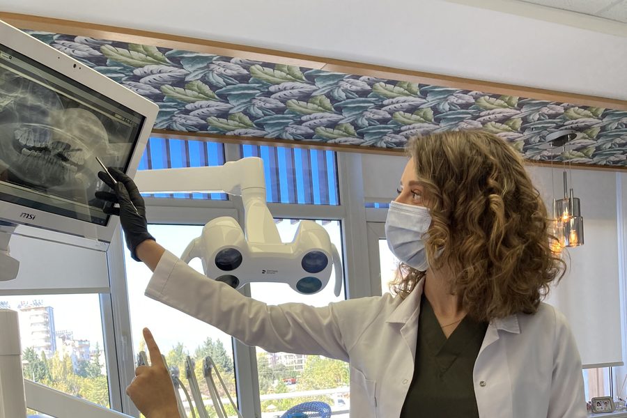
Radiographic and imaging assessment — OPG, CBCT, and MRI as indicated for comprehensive TMJ diagnosis.